

Surgery and equipment
Exoscope vs endoscope: key differences in surgical visualization
A practical comparison between exoscope and endoscope to assess visualization, field access, ergonomics, teaching, and the real needs of the operating room.
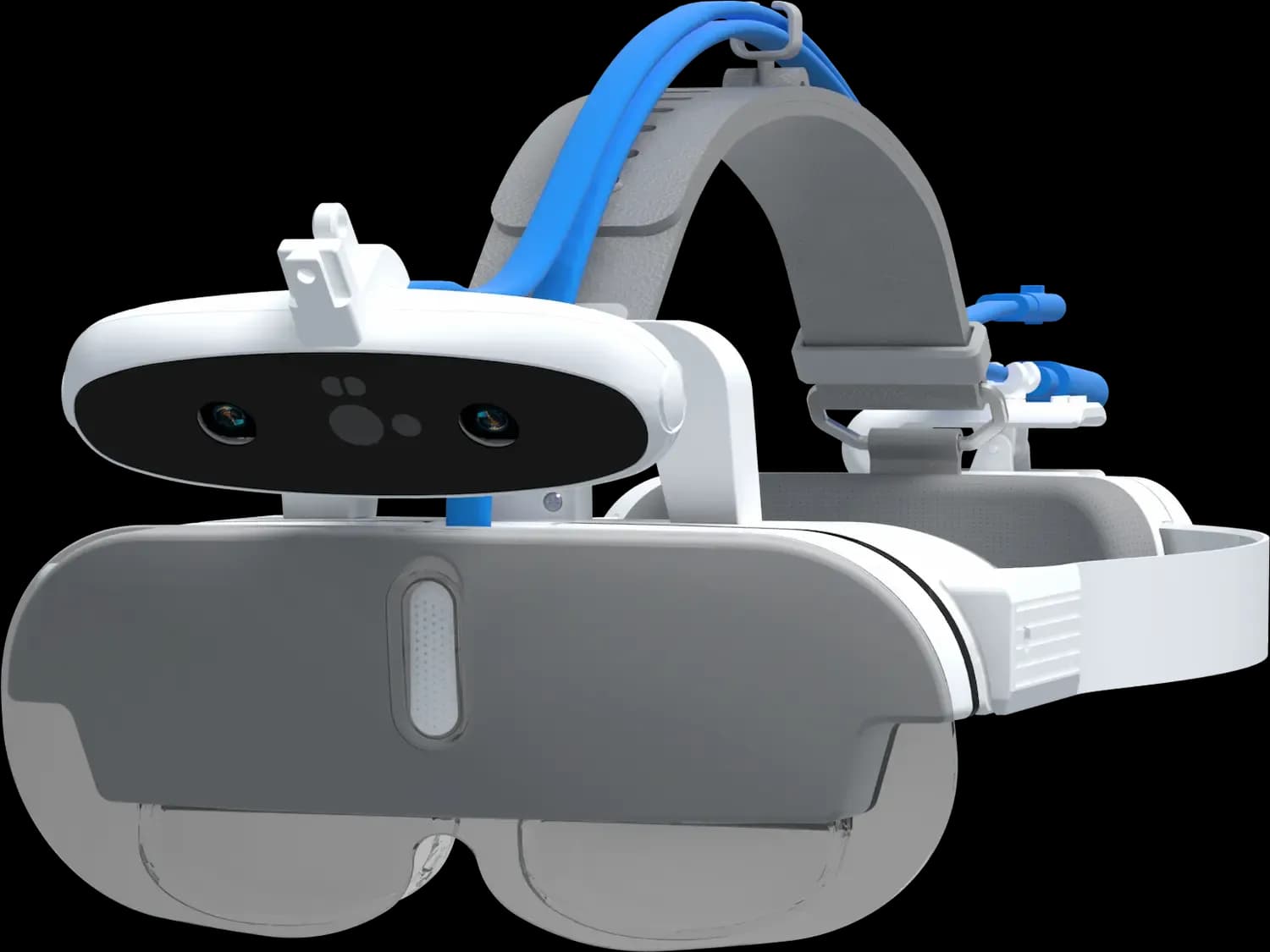
Exoscope vs endoscope describes two different ways of visualizing the surgical field: one external, with the camera positioned outside the patient, and the other internal, using optics introduced through an access route. The choice depends on the procedure, anatomy, available space, and the team’s workflow.
Exoscope vs endoscope: what changes in visualization
The main difference lies in the point from which the image is obtained. In an exoscope, the camera system is placed outside the patient and captures the surgical field from an external position. In an endoscope, the optics are introduced into a cavity, duct, or access pathway to visualize structures from the inside.
This distinction conditions the way surgery is performed. The exoscope is conceptually closer to an external magnified view of the field. The surgeon does not look through eyepieces, but at a screen, and the team can share the same image. The endoscope, by contrast, is associated with procedures in which internal access is part of the surgical approach.
The relationship between image and maneuver also changes. With the exoscope, instruments work in an exposed field or one accessible from the outside. With the endoscope, visualization and instrumentation are coordinated through specific access points, often with a more limited operative space and greater dependence on the optics.
| Criterion | Exoscope | Endoscope |
|---|---|---|
| Camera position | Outside the patient | Introduced through an access route |
| Type of view | Magnified external surgical field | Internal view of cavities or ducts |
| Team workflow | Shared image on a screen | Endoscopic image as the main guide |
| Field access | Exposed field or open/minimally invasive approach depending on the case | Internal access via port, cavity, or tract |
| Key assessment | Ergonomics, screen, working distance, stability | Optics, entry route, orientation, compatible instruments |
Field access: external does not mean equivalent to endoscopic
When comparing exoscope vs endoscope, it is important to avoid a common simplification: this is not about choosing between two interchangeable cameras. Each technology responds to a different way of accessing and working on anatomy.
The endoscope makes it possible to observe internal areas through an entry pathway. That is why it makes sense in procedures in which exploration or intervention is organized around a cavity or a natural or created route. The image depends on the position of the optics, its orientation, and its relationship with the instruments.
The exoscope, on the other hand, is positioned outside the immediate sterile field or integrated via an arm and a camera oriented toward the surgical area. Its usefulness is assessed when the goal is to enlarge and share the view of an external field, without placing the optics inside the patient. This can be especially relevant in environments where the surgeon’s posture, team participation, or procedure documentation carry weight in the decision.
In practice, the question should not be which is better in general, but which visualization method fits the intended approach. If the procedure requires intraluminal or intracavitary vision, the endoscope may be a natural part of the technique. If the field is externally accessible and a shared magnified image is desired, an exoscope may provide a different solution.
Surgical team workflow and ergonomics
Ergonomics is one of the aspects that changes most between the two systems. In traditional optical microscope surgery, the operator is often constrained by eyepieces and fixed positions. With the exoscope, screen-based visualization can allow a working posture less dependent on looking directly through a viewer, provided that the monitor, camera, and table are positioned appropriately.
In endoscopy, the team also works with a screen, but the ergonomic challenge is different. Coordination between the image, the orientation of the optics, and instrument movements requires specific training. In addition, the arrangement of the tower, cables, monitor, and assistants affects the comfort and flow of the procedure.
For the surgical team, the exoscope makes it easier for several people to observe the same image of the external field. This can help coordination, assistance, and teaching, without each professional depending on direct viewing of the field. In endoscopy, shared imaging is also central, but linked to an internal perspective and handling of the optics.
Assessment should include simple but decisive aspects:
- Height and orientation of the main screen.
- Position of the surgeon, assistants, and scrub nurse.
- Available space around the table.
- Stability of the arm or camera support.
- Ease of repositioning the image during the procedure.
- Integration with recording, documentation, or teaching.
At this point, the surgical visualization products from Optimedic Surgery allow you to review options designed for different operating room needs, from imaging systems to complementary solutions.
When an external solution such as MetaSCOPE may fit
An external visualization solution such as MetaSCOPE for surgical visualization may fit when the team is looking for a magnified view of the field without introducing an endoscopic optic. Its design is intended for work with an external camera and screen-based viewing, within a configuration adapted to the operating room.
This can be interesting in procedures where the surgical field is already exposed or accessible from the outside, and where the surgeon and the team need to share a common image. It may also be useful in teaching settings, sessions with observers, or environments where recording and reviewing images are part of the workflow.
However, MetaSCOPE should not be understood as a universal replacement for an endoscope. If the procedure requires internal navigation through cavities, vision through narrow tracts, or a specific optic for an endoscopic technique, the endoscopic approach retains its own logic. The decision depends on the specialty, the procedure, the anatomical field, and the team’s habits.
A prudent way to assess suitability is to ask operational questions:
- Is the field adequately observed from an external position?
- Does the team need everyone to share the same screen image?
- Is the operator’s posture constrained by the current visualization?
- Are recording or teaching common requirements?
- Does the operating room have space for the monitor, arm, and team circulation?
When these answers point to an enlarged external view, it makes sense to request a technical assessment. To do so, the team can request information about MetaSCOPE and review the most suitable configuration for their environment.
Advantages and limitations of each approach
In a comparison of exoscope vs endoscope, advantages should always be interpreted within the clinical and technical context. A system may be suitable for one procedure and impractical for another.
Exoscope
It may add value when:
- Enlarged visualization of an externally accessible field is needed.
- The team wants to share the same screen image.
- Teaching, observation, and documentation are important.
- A reduction in dependence on eyepieces or viewers during work is desired.
- The operating room allows stable placement of the camera, support, and monitor.
It may be less suitable when:
- The required view is intracavitary or intraluminal.
- Access depends on an optic introduced through a port or tract.
- There is not enough space around the table for a comfortable setup.
- The surgical technique is already designed around endoscopy.
Endoscope
It may add value when:
- The procedure requires internal vision through a cavity or access route.
- The technique relies on specific optics and endoscopic equipment.
- The intervention is organized around an intraluminal or intracavitary perspective.
- The team has established endoscopic training and workflow.
It may be less suitable when:
- The field is already exposed and a shared external view is desired.
- The priority is to document or teach an open surgical field from an external perspective.
- Introducing an optic does not add value to the intended approach.
In some departments, the decision is not exclusive. There are operating rooms where different systems coexist depending on the procedure, specialty, and team preference. The key is to define the real use before incorporating technology.
Checklist before incorporating a visualization system
Before deciding between exoscope vs endoscope, it is advisable to carry out a structured review. This checklist helps organize the conversation among surgery, nursing, teaching, purchasing, and technical support.
1. Procedures and approaches
- Which procedures do you want to cover?
- Is the field external, intracavitary, or mixed?
- Does the technique require specific optics?
- Is there a need to change angle frequently?
- Will the system be used in one specialty or several?
2. Operator ergonomics
- How is the surgeon currently positioned?
- Is there sustained cervical flexion, trunk rotation, or forced posture?
- Can the screen be placed on the natural line of sight?
- Does the support allow fine adjustments without interrupting workflow?
- Can assistants maintain a comfortable and safe position?
3. Surgical team and communication
- Who needs to see the image during the procedure?
- Does a shared image improve team coordination?
- Does the scrub nurse or assistant need a secondary screen?
- Is it necessary to integrate the signal with operating room audiovisual systems?
4. Teaching and observation
- Are training sessions or demonstrations held?
- Do residents or students need to see the same image as the operator?
- Does the image need to be projected in the room or sent to another teaching environment?
- Is it important to record procedures for internal review?
5. Recording and documentation
- What type of audiovisual record is needed?
- Is recording part of the department protocol?
- Who manages storage and privacy of the content?
- Is still image capture, video, or both required?
6. Operating room space and logistics
- Is there space for the arm, tower, monitor, or cart?
- Does the system interfere with anesthesia, nursing, or circulation?
- Do cleaning and preparation fit the center’s protocols?
- Can the system be moved between operating rooms if necessary?
7. Training and adoption
- What learning curve will the team have?
- Who will lead standardization of use?
- Is initial training required for surgery and nursing staff?
- How will you assess whether the system fits the department’s routine?
This analysis avoids decisions based solely on the demo image. A visualization system must work in everyday practice, with the procedures, space, and professionals who will actually use it.
Relationship with the surgical microscope
Although this comparison focuses on exoscope vs endoscope, many teams also compare the exoscope with the surgical microscope. The difference is significant: the traditional optical microscope relies on eyepieces, while the exoscope transfers visualization to a screen.
That comparison affects ergonomics, teaching, and field organization. To explore that angle further, you can consult the analysis on digital exoscope vs surgical microscope, focused on the differences between external digital visualization and traditional optical magnification.
Frequently asked questions
Does exoscope vs endoscope always mean choosing one or the other?
Not necessarily. Exoscope vs endoscope compares two visualization approaches, but they can coexist in the same hospital or department. The choice depends on the procedure, anatomical access, the team’s experience, and the purpose of the image: working in a magnified external field or visualizing from the inside.
Can an exoscope replace an endoscope?
It should not be viewed as a universal replacement. An exoscope visualizes from the outside and may fit externally accessible fields. An endoscope allows viewing from inside cavities or access routes. If the technique requires intracavitary vision, the endoscope retains a specific function.
What does MetaSCOPE offer compared with an endoscopic approach?
MetaSCOPE can provide external screen-based visualization for procedures where the field is viewed from the outside and the image needs to be shared with the team. Its suitability should be assessed according to specialty, ergonomics, teaching, recording, and available space, without assuming that it replaces endoscopic techniques.
What should an operating room review before incorporating an exoscope?
It should review the intended procedures, operator posture, monitor placement, space around the table, recording needs, teaching, and team training. It is also advisable to carry out a practical evaluation with the professionals who will use the system under conditions similar to usual practice.
Next step to assess surgical visualization
The comparison between exoscope and endoscope should begin with the surgical approach, not only with the technology. If the goal is to evaluate an external solution such as MetaSCOPE, the sensible next step is to review procedures, ergonomics, and audiovisual requirements with a specialized technician.
To move forward, the team can request a MetaSCOPE assessment and compare the configuration with the real needs of the operating room.